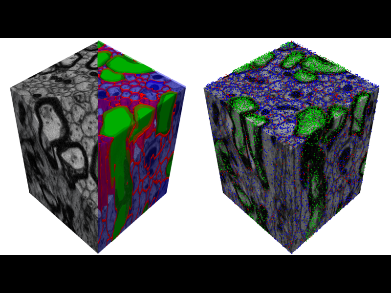
 Segmentation of 3D electron microscopy (SBFSEM) data to perform realistic simulations of the diffusion process in the brain.<= more info =>
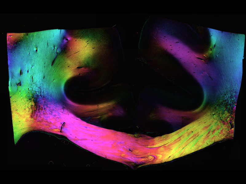 Unraveling the mesoscopic structure of the human corpus callosum with polarized light imaging.<= more info =>
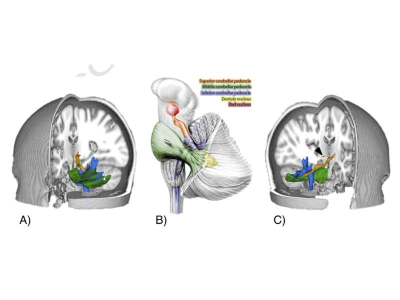 Creating an atlas of the cerebellar peduncles with tractography in the Human Connectome Project dataset.<= more info =>
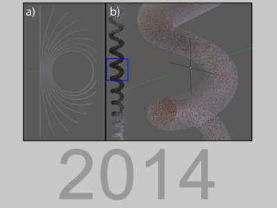 Investigating the mechanisms of diffusion restriction along axons with MRI, simulations and polarized light imaging.<= more info =>
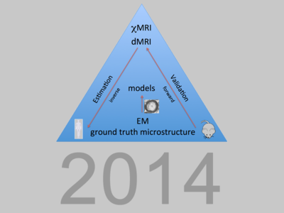 in this project, I'm developing MR methods to characterize plasticity-induced white matter changes<= more info =>
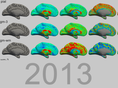 a project to characterize the effects of cortical folding and layering on diffusion tensor measures in the gyri of the medial cortical areas<= more info =>
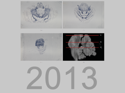 comparing ex vivo MR scans of the cerebellum to volumetrically-reconstructed histologically-stained serial sections of the same specimen<= more info =>
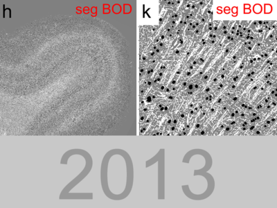 validation of the biophysical diffusion model NODDI and investigations into fibre insertion patterns in the cortex<= more info =>
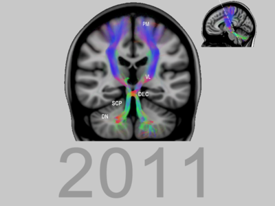 creating neuroanatomy teaching tools: plastinated white matter dissection specimens, combined with tractography images<= more info =>
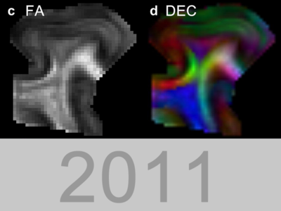 identifying cortical layers (e.g. the line of Gennari) with diffusion imaging in human tissue samples of the primary visual cortex<= more info =>
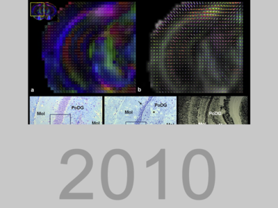 in an Alzheimer mouse model, abnormalities in diffusion images were demonstrated in white and grey matter (hippocampus)<= more info =>
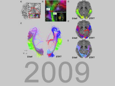 development of a tractography method that improves tracking by incorporating high resolution structural T2*-weighted images<= more info =>
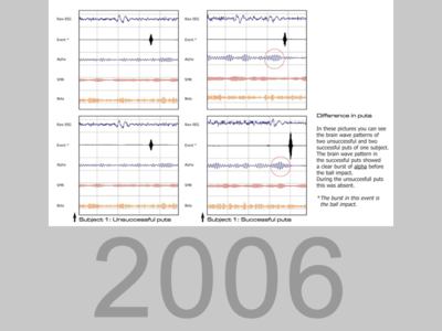 methods development for sports training (e.g. golf) by identifying an optimal EEG-signature and providing neurofeedback to train the pattern<= more info =>
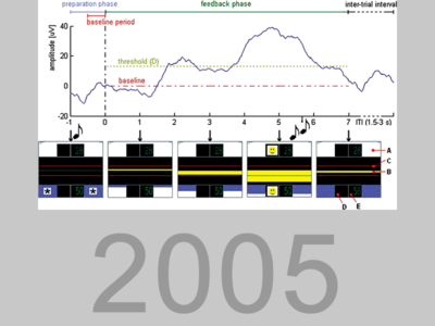 building a brain-computer interface operated by EEG-signals: the sensorimotor rhythm and slow cortical potential<= more info =>
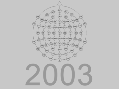 investigating the relation between sampling rate and localization of epileptic foci using a high-end EEG system<= more info =>
 hardware and software development for a scanning stage used in photoacoustic mammography to detect and monitor breast cancer without using x-rays<= more info =>
3d electron microscopy, fmrib 2014
▲back to toppolarized light imaging, fmrib 2014
▲back to topcerebellar white matter atlas, fmrib 2014
▲back to topAbstract
Imaging of the cerebellar cortex, deep cerebellar nuclei and their connectivity are gaining attraction, due to the important role the cerebellum plays in cognition and motor control. Atlases of the cerebellar cortex and nuclei are used to locate regions of interest in clinical and neuroscience studies. However, the white matter that connects these relay stations is of at least similar functional importance. Damage to these cerebellar white matter tracts may lead to serious language, cognitive and emotional disturbances, although the pathophysiological mechanism behind it is still debated. Differences in white matter integrity between patients and controls might shed light on structure-function correlations. A probabilistic parcellation atlas of the cerebellar white matter would help these studies by facilitating automatic segmentation of the cerebellar peduncles, the localization of lesions and the comparison of white matter integrity between patients and controls.
In this work a digital three-dimensional probabilistic atlas of the cerebellar white matter is presented, based on high quality 3T, 1.25mm resolution diffusion MRI data from 90 subjects participating in the Human Connectome Project. The white matter tracts were estimated using probabilistic tractography. Results over 90 subjects were symmetrical and trajectories of superior, middle and inferior cerebellar peduncles resembled the anatomy as known from anatomical studies.
This atlas will contribute to a better understanding of cerebellar white matter architecture. It may eventually aid in defining structure-function correlations in patients with cerebellar disorders.
van Baarsen K.M., Kleinnijenhuis M., Jbabdi S., Sotiropoulos S.N., Grotenhuis, J.A., van Cappellen van Walsum A.M. (2015). A probabilistic atlas of the cerebellar white matter. NeuroImage, in press. doi:10.1016/j.neuroimage.2015.09.014
diffusion restriction along axons, fmrib 2014
▲back to topMicrostructure imaging relies on restriction of diffusion by cell components (such as the outer cell membrane). The occurence of restriction is well known for the direction perpendicular to axons. This has been used to derive useful microstructural parameters like axon diameter distributions and volume fractions. In this project, we investigate restrictions in the direction of axonal bundles. It is often thought that diffusion is relatively free or independent of diffusion time in this direction. However, at long diffusion times, it becomes apparent that restrictions also occur in the direction of the corpus callosum: the fibre tract that is thought to be most coherently organized.
We investigate this phenomenon using a set of different tools. In vivo and post mortem diffusion MRI are used to characterize the diffusion behaviour, while Monte Carlo simulations and Polarized Light Imaging are used to identify and evaluate potential mechanisms.
Kleinnijenhuis, M., Mollink, J., Kinchesh, P., Lam, W.W., Galinsky, V.L., Frank, L.R., Smart, S.C., Jbabdi, S., & Miller, K.L. (2015). Monte Carlo diffusion simulations disambiguate the biophysical mechanisms of diffusion hinderance along tracts. Proceedings of the International Society for Magnetic Resonance in Medicine. Toronto, Canada.
Mollink, J., Kleinnijenhuis, M., Sotiropoulos, S.N., Ansorge, O., Jbabdi, S., & Miller, K.L. (2015). Diffusion restriction along fibres: How coherent is the corpus callosum? Proceedings of the International Society for Magnetic Resonance in Medicine. Toronto, Canada.
MR methods for plasticity, fmrib 2014
▲back to topIt has long been thought that human brain structure is invariable in adulthood. Research over the past decade, however, suggests that there is considerable structural plasticity, i.e. neuronal reorganisation, in the adult brain. Using magnetic resonance imaging (MRI), this can be detected in grey matter as well as white matter, where an important constituent—myelin—appears to change when learning a new skill.
Yet, it cannot be discerned which changes in neuron microstructure (e.g. myelin thickness, axon diameter, packing efficiency) cause these subtle variations in MRI contrast. Crucially, lacking these microstructural MRI markers holds back development of treatment approaches (e.g. in stroke and multiple sclerosis) and prevention strategies for neurodegenerative disease.
This project aims to develop these MRI methods for estimating quantities specific to microstructural components. A significant part of the project is concerned with the novel approach of using ultrastructural measurements from electron microscopy as the basis for these models. Application to fundamental questions about plasticity (does the brain grow new myelin when learning?) in animal models and humans will assess the validity and benefit of our approach to the measurement of microstructure with MRI.
DTI of gyrencephaly, donders 2013
▲back to topAbstract
Gyrification of the human cerebral cortex allows many more neurons as compared to other mammals. For neuroimaging, however, gyrification complicates analysis, e.g. cortical laminae occupy different depths in gyri and sulci. Recently, diffusion-weighted imaging has explored cortical fibre architecture. This warrants investigating the relation between diffusion tensors and gyral patterns.
High-resolution (1mm3) diffusion-weighted imaging of the hemispheres’ medial walls was performed at 7T. Data were resampled to surfaces in cortex and underlying white matter. Cortical surfaces obeyed the equivolume principle for laminae over cortical curvature. Tensor metrics were averaged over curvature bins to map characteristic patterns in the gyrus.
Diffusivity, anisotropy and radiality varied with curvature and over laminae. Radiality was maximal in intermediate laminae near the gyral crown, not in white matter or on the crown. In the fundus, deep layers had tangential tensor orientations. In white matter, tensors changed from radial under the crown to tangential under the bank/fundus. White matter anisotropy increased from crown to fundus.
This characteristic architecture is in accordance with ex vivo diffusion MR-microscopy and histological studies. Results indicate the necessity of taking into account the gyral pattern for cortical diffusion analysis. Additionally, data suggest a tractography bias towards the crown.
Kleinnijenhuis M., van Mourik T., Norris D.G., Ruiter D.J., van Cappellen van Walsum A-M., Barth M. (2015) Diffusion tensor characteristics of gyrencephaly using high resolution diffusion tensor imaging in vivo at 7T. NeuroImage, 109, 378-387. doi:10.1016/j.neuroimage.2015.01.001
tractography validation, anatomy radboudumc 2013
▲back to topAbstract
Probabilistic tractography allows for reconstruction of white matter fibers in the brain using diffusion weighted magnetic resonance imaging (DWI) and is a tool for studying connectivity in the brain. For research and clinical application of tractography it is essential to know whether tract reconstructions correspond with their anatomical position. The gold standard for validation of tractography is the comparison with histological fiber staining in post mortem tissue. In this study probabilistic tractography is applied to reconstruct the dentaterubrothalamic tract (DRT) and validated with a myelin stain.
DWI data were acquired with a 7T MRI scanner from an ex vivo human brain specimen. Probabilistic tractography was performed with FSL. Subsequently, the cerebellum was prepared for histological processing and cut into sections which were stained for myelin. The sections were digitized for 3D reconstruction. Interslice alignment was performed to align neighbouring slices with each other prior to 3D reconstruction resulting in the histological volume. The DRT was segmented in the histological volume which served as validation reference. Tractography and the validation reference were registered with each other for voxelwise comparison. ROC analysis, similarity index (SI) and miss fraction (MF) were computed for several tract probability thresholds for validation.
ROC analysis resulted in a sensitivity of 0.8348 with a specificity of 0.9963. Optimal SI of 0.63 and 0.78 with a MF of 0.37 and 0.17 were found for the whole tract and the superior cerebellar peduncle (SCP) region, respectively. In total, 85% of the reference tract was located within 1 mm range from the border of tractography.
The feasibility of histology-based validation of the diffusion weighted tractography in the DRT has been demonstrated in this study. A plausible 3D reconstruction of the DRT was achieved by histological sectioning as well as probabilistic tractography. SI reported here have been regarded as excellent in other segmentation studies. Spatial alignment between tractography and the histology based tract reaches an accuracy which is ready for comparison to in vivo studies of the DRT with probabilistic tractography.
Mollink J., van Baarsen K.M., Dederen P.J.W.C., Foxley S., Miller K.L., Jbabdi S., Slump C.H., Grotenhuis J.A., Kleinnijenhuis M., van Cappellen van Walsum A.M. (2015). Dentatorubrothalamic tract localization with post mortem MR diffusion tractography compared to histological 3D reconstruction. Brain Structure and Function, in press.
Mollink, J., van Baarsen, K., Slump, C.H., Foxley, S., Miller, K.L., Norris, D.G., Kleinnijenhuis, M., & van Cappellen van Walsum, A-M. (2014). Validation of diffusion weighted tractography in the dentatorubrothalamic tract. Organization for Human Brain Mapping. Hamburg, Germany, 2014.
Mollink, J., van Baarsen, K., Slump, C.H., Foxley, S., Miller, K.L., Norris, D.G., Kleinnijenhuis, M., & van Cappellen van Walsum, A-M. (2014). Validation of diffusion weighted tractography in the dentatorubrothalamic tract. Nederlandse Anatomenvereniging. Lunteren, The Netherlands, 2014.
Sikma, K.-J., Kleinnijenhuis, M., Barth, M., Dederen, P. J., Kuesters, B., Slump, C. H., & Van Cappellen van Walsum, A.-M. (2010). Validating tractography from DWI/SWI data with 3D reconstructed histological data of post-mortem human brain tissue. Nederlandse Vereniging voor Technische Geneeskunde. Enschede, The Netherlands.
gyral fibre architecture, anatomy radboudumc 2012
▲back to topAbstract
Recent advances in diffusion MRI methods allow imaging of fibres in the gyri of the cerebral hemispheres. High resolution promises to improve structural connectomics by enabling tractography over the gray-white matter boundary. The study of disease processes can be promoted by microstructural markers from biophysical models.
To validate and explore these new possibilities, ex vivo diffusion MRI of human gyral samples from the primary visual cortex was performed using a multi-shell protocol. The NODDI model was used to derive neurite volume fraction and neurite orientation dispersion. The neurite dispersion and density were compared to histological data (nucleus, myelin and neurofilament stains) by volume fraction and structure tensor analysis.
The MR-derived neurite volume fraction was 0.8 for gyral white matter. In the cortex, neurite density was layer-specific: 0.37 for the stria of Gennari; 0.25 for supragranular layers; 0.34 for infragranular layers. Myelin histology showed higher correlation with MR-derived neurite volume fraction than the neurofilament stain. NODDI dispersion maps showed additional lamination in infragranular layers (the inner band of Baillarger). At the gray-white matter boundary, a layer of high dispersion is found lining the cortical sheet. Histology indicated that the fibre configurations varied from crossing on the gyral banks to fanning near the gyral crowns.
The NODDI model has been shown to generate plausible estimates of fibre properties in the gyral white and gray matter. The histology indicated that models to be used for diffusion imaging of gyral fibre configurations should be able to model crossing as well as fanning configurations.
Tariq, M., Kleinnijenhuis, M., van Cappellen van Walsum, A.-M., & Zhang, H. (2015). Validation of NODDI estimation of dispersion anisotropy in V1 of the human neocortex. Proceedings of the International Society for Magnetic Resonance in Medicine. Toronto, Canada.
Kleinnijenhuis M., Zhang H., Wiedermann D., Küsters B., Dederen P.J., Norris D.G., van Cappellen van Walsum A-M. (in preparation). Fibres in the gyrus: characteristics in ex vivo MRI and histology.
Kleinnijenhuis, M., Zhang, H., Wiedermann, D., Norris, D. G. & Van Cappellen van Walsum, A.-M. (2013). Detailed laminar characteristics of the human neocortex revealed by NODDI. International Conference on Magnetic Resonance Microscopy Cambridge, United Kingdom.
Kleinnijenhuis, M., Zhang, H., Wiedermann, D., Kuesters, B. Norris, D. G., & Van Cappellen van Walsum, A.-M. (2013). Detailed laminar characteristics of the human neocortex revealed by NODDI and histology. Organization for Human Brain Mapping (p. 1685). Seattle, United States of America.
Kleinnijenhuis, M., Zhang, H., Wiedermann, D., Kuesters, B., Van Cappellen van Walsum, A.-M., & Norris, D. G. (2013). Detailed laminar characteristics of the human neocortex revealed by NODDI. Proceedings of the International Society for Magnetic Resonance in Medicine (p. 5100). Salt Lake City, United States of America.
dissection and plastination for education, anatomy radboudumc 2011
▲back to topAbstract
In recent years, there has been a growing interest in white matter anatomy of the human brain. With advances in brain imaging techniques, the significance of white matter integrity for brain function has been demonstrated in various neurological and psychiatric disorders. As the demand for interpretation of clinical and imaging data on white matter increases, the needs for white matter anatomy education are changing. Because cross-sectional images and formalin-fixed brain specimens are often insufficient in visualizing the complexity of three-dimensional (3D) white matter anatomy, obtaining a comprehensible conception of fiber tract morphology can be difficult. Fiber dissection is a technique that allows isolation of whole fiber pathways, revealing 3D structural and functional relationships of white matter in the human brain.
In this study, we describe the use of fiber dissection in combination with plastination to obtain durable and easy to use 3D white matter specimens that do not require special care or conditions. The specimens can be used as a tool in teaching white matter anatomy and structural connectivity. We included four human brains and show a series of white matter specimens of both cerebrum and cerebellum focusing on the cerebellar nuclei and associated white matter tracts, as these are especially difficult to visualize in two-dimensional specimens and demonstrate preservation of detailed human anatomy. Finally, we describe how the integration of white matter specimens with radiological information of new brain imaging techniques such as diffusion tensor imaging tractography can be used in teaching modern neuroanatomy with emphasis on structural connectivity.
Arnts, H., Kleinnijenhuis, M., Kooloos, J., Schepens-Franke, A., & Van Cappellen van Walsum, A.-M. (2014). Combining fiber dissection, plastination, and tractography for neuroanatomical education: Revealing the cerebellar nuclei and their white matter connections. Anatomical Sciences Education, 7(1), 47-55. doi:10.1002/ase.1385
diffusion MR microscopy of the neocortex, anatomy radboudumc 2011
▲back to topAbstract
One of the most prominent characteristics of the human neocortex is its laminated structure. The first person to observe this was Francesco Gennari in the second half the 18th century: in the middle of the depth of primary visual cortex, myelinated fibres are so abundant that he could observe them with bare eyes as a white line. Because of its saliency, the stria of Gennari has a rich history in cyto- and myeloarchitectural research as well as in magnetic resonance (MR) microscopy.
In the present paper we show for the first time the layered structure of the human neocortex with ex vivo diffusion weighted imaging (DWI). To achieve the necessary spatial and angular resolution, primary visual cortex samples were scanned on an 11.7 T small-animal MR system to characterize the diffusion properties of the cortical laminae and the stria of Gennari in particular.
The results demonstrated that fractional anisotropy varied over cortical depth, showing reduced anisotropy in the stria of Gennari, the inner band of Baillarger and the deepest layer of the cortex. Orientation density functions showed multiple components in the stria of Gennari and deeper layers of the cortex. Potential applications of layer-specific diffusion imaging include characterization of clinical abnormalities, cortical mapping and (intra)cortical tractography.
We conclude that future high-resolution in vivo cortical DWI investigations should take into account the layer-specificity of the diffusion properties.
Kleinnijenhuis, M., Zerbi, V., Kuesters, B., Slump, C. H., Barth, M., & Van Cappellen van Walsum, A.-M. (2013). Layer-specific diffusion weighted imaging in human primary visual cortex in vitro. Cortex, 49(9), 2569-2582. doi:10.1016/j.cortex.2012.11.015
Kleinnijenhuis, M., Barth, M., Zerbi, V., Sikma, K.-J., Kuesters, B., Slump, C. H., Norris, D. G., Ruiter, D.J., & Van Cappellen van Walsum, A.-M. (2012). A new perspective on the cortical laminar pattern of the primary visual cortex in the occipital lobes. International School of Clinical Neuroanatomy: the Clinical Neuroanatomy of the Occipital Lobes. Ragusa, Italy.
Kleinnijenhuis, M., Barth, M., Zerbi, V., Sikma, K.-J., Kuesters, B., Slump, C. H., Norris, D. G., Ruiter, D.J., & Van Cappellen van Walsum, A.-M. (2011). In vitro layer-specific Diffusion Weighted Imaging in human primary visual cortex. Nederlandse Vereniging voor Technische Geneeskunde. Woerden, The Netherlands.
Kleinnijenhuis, M., Barth, M., Zerbi, V., Sikma, K.-J., Kuesters, B., Slump, C. H., Norris, D. G., Ruiter, D.J., & Van Cappellen van Walsum, A.-M. (2011). In vitro layer-specific Diffusion Weighted Imaging in human primary visual cortex. Brain Circuitry and its Disorders. Doorwerth, The Netherlands.
Kleinnijenhuis, M., Barth, M., Zerbi, V., Sikma, K.-J., Kuesters, B., Slump, C. H., Norris, D. G., Ruiter, D.J., & Van Cappellen van Walsum, A.-M. (2011). Layer-specific diffusion weighted imaging in human primary visual cortex in vitro. Organization for Human Brain Mapping (p. 2509). Quebec City, Canada.
Kleinnijenhuis, M., Sikma, K.-J., Barth, M., Dederen, P. J., Zerbi, V., Kuesters, B., Ruiter, D. J., Slump, C. H., & van Cappellen van Walsum, A.-M. (2011). Validation of Diffusion Weighted Imaging of cortical anisotropy by means of a histological stain for myelin. Proceedings of the International Society for Magnetic Resonance in Medicine (p. 2240).
Kleinnijenhuis, M., Sikma, K.-J., Barth, M., Dederen, P. J., Zerbi, V., Kuesters, B., Ruiter, D.J., Slump, C. H., & van Cappellen van Walsum, A.-M. (2011). Validation of Diffusion Weighted Imaging of cortical anisotropy by means of a histological stain for myelin. Proceedings of the International Society for Magnetic Resonance in Medicine: Benelux chapter.
mouse brain diffusion imaging, anatomy radboudumc 2010
▲back to topAbstract
In patients with Alzheimer's disease (AD) the severity of white matter degeneration correlates with the clinical symptoms of the disease.
In this study, we performed diffusion-tensor magnetic resonance imaging at ultra-high field in a mouse model for AD (APPswe/PS1dE9) in combination with a voxel-based approach and tractography to detect changes in water diffusivity in white and gray matter, because these reflect structural alterations in neural tissue.
We found substantial changes in water diffusion parallel and perpendicular to axonal tracts in several white matter regions like corpus callosum and fimbria of the hippocampus, that match with previous findings of axonal disconnection and myelin degradation in AD patients. Moreover, we found a significant increase in diffusivity in specific hippocampal subregions, which is supported by neuronal loss as visualized with Klüver-Barrera staining.
This work demonstrates the potential of ultra-high field diffusion-tensor magnetic resonance imaging as a noninvasive modality to describe white and gray matter structural changes in mouse models for neurodegenerative disorders, and provides valuable knowledge to assess future AD prevention strategies in translational research.
Zerbi, V.*, Kleinnijenhuis, M.*, Fang, X., Jansen, D., Veltien, A., Van Asten, J., Timmer, N., et al. (2012). Gray and white matter degeneration revealed by diffusion in an Alzheimer mouse model. Neurobiology of aging, 34(5), 1440-1450. doi:10.1016/j.neurobiolaging.2012.11.017 *equal contribution
structure tensor informed fibre tractography (STIFT), donders 2009
▲back to topAbstract
Structural connectivity research in the human brain in vivo relies heavily on fiber tractography in diffusion-weighted MRI (DWI). The accurate mapping of white matter pathways would gain from images with a higher resolution than the typical ~ 2 mm isotropic DWI voxel size. Recently, high field gradient echo MRI (GE) has attracted considerable attention for its detailed anatomical contrast even within the white and gray matter. Susceptibility differences between various fiber bundles give a contrast that might provide a useful representation of white matter architecture complementary to that offered by DWI.
In this paper, Structure Tensor Informed Fiber Tractography (STIFT) is proposed as a method to combine DWI and GE. A data-adaptive structure tensor is calculated from the GE image to describe the morphology of fiber bundles. The structure tensor is incorporated in a tractography algorithm to modify the DWI-based tracking direction according to the contrast in the GE image.
This GE structure tensor was shown to be informative for tractography. From closely spaced seedpoints (0.5 mm) on both sides of the border of 1) the optic radiation and inferior longitudinal fasciculus 2) the cingulum and corpus callosum, STIFT fiber bundles were clearly separated in white matter and terminated in the anatomically correct areas. Reconstruction of the optic radiation with STIFT showed a larger anterior extent of Meyer's loop compared to a standard tractography alternative. STIFT in multifiber voxels yielded a reduction in crossing-over of streamlines from the cingulum to the adjacent corpus callosum, while tracking through the fiber crossings of the centrum semiovale was unaffected.
The STIFT method improves the anatomical accuracy of tractography of various fiber tracts, such as the optic radiation and cingulum. Furthermore, it has been demonstrated that STIFT can differentiate between kissing and crossing fiber configurations. Future investigations are required to establish the applicability in more white matter pathways.
Kleinnijenhuis, M., Barth, M., Alexander, D. C., Van Cappellen van Walsum, A.-M., & Norris, D. G. (2012). Structure Tensor Informed Fiber Tractography (STIFT) by combining gradient echo MRI and diffusion weighted imaging. NeuroImage, 59(4), 3941-54. doi:10.1016/j.neuroimage.2011.10.078
real-life neurofeedback, brainquiry 2006
▲back to topAfter the BCI study for my MSc, I started a job at Brainquiry where I investigated performance improvement by brain wave training. In tasks that require a great deal of concentration, it can be a great feeling to be “in the zone”: everything seems to be going extremely well without any effort. We wondered 1) whether this “in the zone” feeling is reflected in the EEG, 2) what this might look like, and importantly, 3) whether performance could be improved by letting you know whether your brain is “in the zone”.
This definitely called for a field study. Golf was the sport that had the edge. We recorded EEG from golfers while putting and scored the puts as hit or miss. In the analysis, we separated hits from misses and looked at the EEG signals in the seconds before the put. With event-locked averaging of frequency bands, we found that there’s no one specific characteristic EEG pattern that predicts success. Rather, this seemed to be quite variable between individuals. Some people showed high Alpha activity just before successful puts, but others had different ‘preferred’ frequencies. In some people we even found evidence that their brain had timing issues on their bad puts.
In itself, this personalized optimal EEG profile for good performance was already a pretty interesting observation. But we took it a step further. We incorporated this information in the training routine of golfers by only allowing putting when their brain waves were ‘just right’ for them—a sorta Goldielocks brain state. Let me tell you: they weren’t very happy about this (golfers, as any sportsperson, don’t really like you to mess with their routine). But it worked!
Feedback improved their putting accuracy. In runs with feedback on their brain state, performance increased compared to the baseline run without feedback. In a third run without feedback, performance dropped again, to increase once more when signal was turned on again in a fourth run. After the experiment in amateur golfers, we extended the approach to professionals, as well as to other sports (shooting, darts). When all of this went reasonably well, we even made a product out of it. Still waiting for this to make me rich ;)
Fallahpour, K., Kleinnijenhuis, M., Arns, M.W. (2011). Peak Performance Using Personalized and Task-Specific Event-Locked EEG: A Wireless, Personalized Medicine Approach to Sports Performance Enhancement. In: Strack BW, Linden MK, Wilson VS (eds). Biofeedback & Neurofeedback Applications in Sport Psychology. Association for Applied Psychophysiology and Biofeedback.
Arns, M., Kleinnijenhuis, M., Fallahpour, K., & Breteler, R. (2008). Golf Performance Enhancement and Real-Life Neurofeedback Training Using Personalized Event-Locked EEG Profiles. Journal of Neurotherapy, 11(4), 11-18. doi:10.1080/10874200802149656
Kleinnijenhuis, M., Arns, M. W., & Rijpma, J. (2006). Golf performance enhancement by means of real-life neurofeedback training based on personalized event-locked EEG profiles. In E. Peper & M. Fuhs (Eds.), Applied Psychophysiology and Biofeedback: Abstracts of Scientific Papers Presented at the 10th Anniversary Meeting of the Biofeedback Foundation of Europe (Vol. 31, pp. 346-347). Vienna, Austria.
brain-computer interfacing, brainquiry 2005
▲back to topFor my MSc thesis, I developed a brain-computer interface (BCI)—or as it is stated in the title of my thesis: a “discrete-trial based biofeedback system”. This was back in the days when not a lot of people worked on BCI. At the company where I did this assignment (Brainquiry), portable devices for a few channels EEG were developed. This is of course great for brain-computer interfacing as you can easily take you research to the field (see also real-life neurofeedback).
The experiment we did used one of three signals detected by this device—two different EEG frequencies and a skin conductance measure (GSR). The brain signals were the sensori-motor rhytm (SMR) and the slow cortical potential (SCP). Both already were known to be controllable by operant conditioning. For SMR, the main application was epilepsy treatment, while the SCP had been used in classic BCI research in ALS patients. The new elements in my project were bidirectional training and a direct comparison between the two techniques. To get the experimental design we wanted compatible with the EEG equipment, I developed an elaborate design in BioExplorer. If you happen to have compatible equipment, it’s available here.
The outcome of the experiment is line with what many BCI studies find: there’s a large variability in the extent to which people are able to control their brain activity. Furthermore, it was found that it’s important to shape the response by manipulating the difficulty of the task. We also had some insight into how the signals are related to each other (as all three signals can be thought to reflect arousal). Two papers resulted from this study, one on SMRvsSCP and one on SCPvsGSR, as well as my MSc thesis.
Spronk, D., Kleinnijenhuis, M., Van Luijtelaar, G., & Arns, M. (2010). Discrete-Trial SCP and GSR Training and the Interrelationship Between Central and Peripheral Arousal. Journal of Neurotherapy, 14(3), 217-228. doi:10.1080/10874208.2010.501501
Kleinnijenhuis, M., Arns, M., Spronk, D., Breteler, R., & Duysens, J. (2008). Comparison of Discrete-Trial-Based SMR and SCP Training and the Interrelationship Between SCP and SMR Networks: Implications for Brain-Computer Interfaces and Neurofeedback. Journal of Neurotherapy, 11(4), 19-35. doi:10.1080/10874200802162808
Kleinnijenhuis, M. (2007). Comparison of SMR and SCP training employing a newly developed discrete-trial based biofeedback system: Implications for Brain-Computer Interfaces, Neurofeedback and the interrelationship between SCP and SMR networks. MSc Thesis, Radboud University Nijmegen, Nijmegen, The Netherlands.
Breteler, R., Kleinnijenhuis, M., Spronk, D., Heesen, E., & Arns, M. (2006). The acquisition of control in self-regulation of Galvanic skin response and slow cortical potentials: A randomized trial. In E. Peper & M. Fuhs (Eds.), Applied Psychophysiology and Biofeedback: Abstracts of Scientific Papers Presented at the 10th Anniversary Meeting of the Biofeedback Foundation of Europe (pp. 354-355). Vienna, Austria.
high definition encephalography, mst 2003
▲back to topIn 2003, I did my BSc internship at the Medisch Spectrum Twente hospital in Enschede. The department of Clinical Neurophysiology just bought a new EEG system with 64 channels: very impressive at the time. Apart from general implementation of the system, during my time there I have specifically looked at the relationship between sampling rate and source localization of epileptic foci.
photoacoustic mammography, ut 2002
▲back to topAt the University of Twente I have worked on a project on photoacoustic mammography. In photoacoustic imaging short pulses of light are delivered to tissue. The light is partly absorbed and causes a rapid expansion of the tissue. When tissue expands, ultrasound waves are emitted and are conducted through the tissue. This can be picked up outside the body by ultrasound detectors. Because the light at the wavelengths used is absorbed particularly well by the hemoglobin in the blood, the photoacoustic signal is different for highly vascularized tumors than for healthy breast tissue. This is a promising technique for detecting and monitoring breast cancer without using X-rays.
Link to the University of Twente’s BMPI group.


