|
 |
|
|
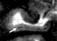
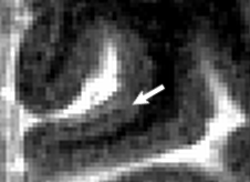
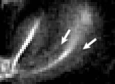
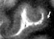
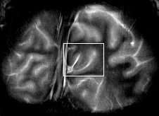
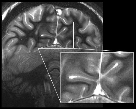
The striate cortex, responsible for the first level processing visual signals, is so named because of the myelinated stripe (layer IVb) within the cortex. This layer is less than 500 um thick so to image this it has been necessary to combine optimised MR contrast with an in-plane resolution of 300 um x 300 um. The images shown above were obtained in a scan lasting just under 30 minutes. Imaging myeloarchitecture in vivo is important since it may provide a link between functional imaging methods and classical neuroscience which describes the functional segregation of the cortex in term of cortical structure. (See Clare, S., Jezzard, P., Matthews, P.M. (2002) Identification of the myelinated layers in striate cortex on high resolution MRI at 3 Tesla. Proc. 10th Meeting International Society for Magnetic Resonance in Medicine, 1465.)